-
×
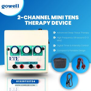 Ultimate Pain Relief with Gowell 2-Channel Mini TENS Machine: Fast & Effective Muscle Stimulation
1 × ৳ 14,000.00
Ultimate Pain Relief with Gowell 2-Channel Mini TENS Machine: Fast & Effective Muscle Stimulation
1 × ৳ 14,000.00
Subtotal: ৳ 14,000.00
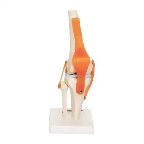
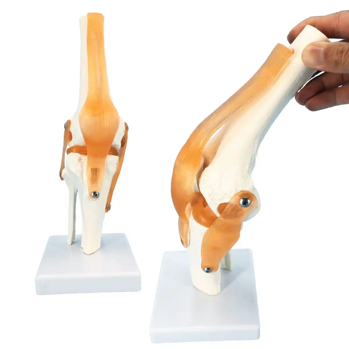
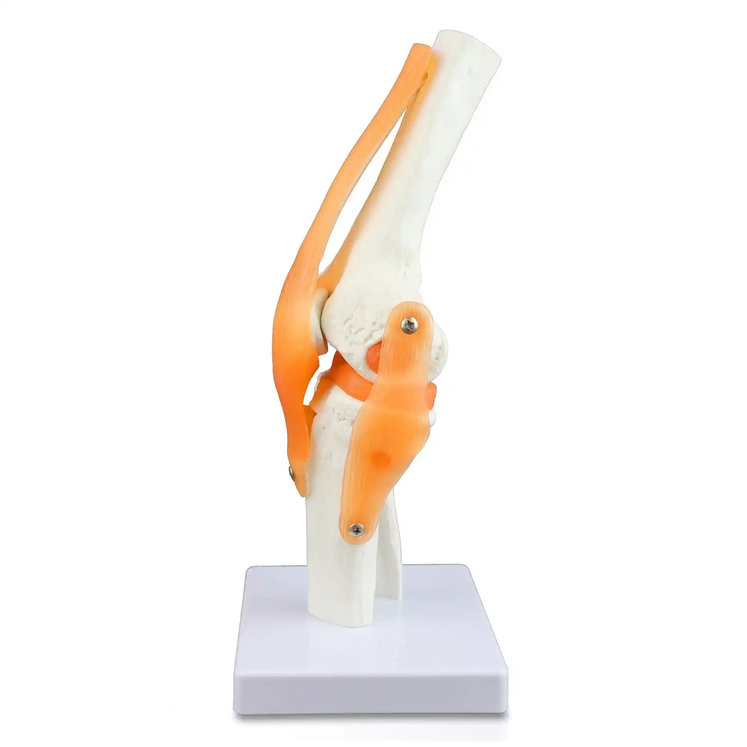
Product Name: Human Knee Joint Anatomical Model
Material: High-quality plastic or resin for durability and detail
Size: Standard adult-sized knee joint model
Components: Includes femur, tibia, patella, ligaments, menisci, and tendons
Color: Natural bone-like color
Base: Sturdy stand for easy display and use
Dimensions: 10 x 8 x 12 inches
Weight: Approx. 1.5 kg
Usage: Educational, patient education, and surgical planning
Looking to understand knee anatomy better? The Human Knee Joint Anatomical Model offers a detailed, realistic representation of the bones, ligaments, tendons, and menisci. Perfect for medical students, orthopedic surgeons, and patient education, it provides an up-close view of knee structure and movement. Order now for a clearer understanding of knee health!
Looking to learn more about the knee joint or educate others? The Human Knee Joint Anatomical Model is a must-have tool for medical students, physical therapists, and anyone interested in knee anatomy. This highly detailed model gives you an up-close look at the bones, ligaments, menisci, and more, offering a deeper understanding of the knee’s structure and function.
Detailed Representation: The Human Knee Joint Anatomical Model includes all essential parts like the femur, tibia, patella, ligaments, tendons, and menisci. Every detail is replicated for a realistic, functional understanding of the knee joint.
Articulating Joints: The model allows for flexion and extension, showing the knee’s natural movements. You can visualize how the knee joint moves in different directions, providing an in-depth look at its mechanics.
Ligaments and Tendons: The model highlights important ligaments of the knee joint such as the ACL, PCL, MCL, and LCL, which are crucial for stabilizing the knee. It also features patellar tendons for added realism.
Menisci and Synovial Membrane: The model showcases the medial and lateral menisci, helping users understand their role in shock absorption. It also includes the synovial membrane and fluid for a complete representation.
Durable Construction: Made from high-quality materials, the Human Knee Joint Anatomical Model is designed for long-term use in educational environments, medical practices, and clinics.
Hands-On Learning: This anatomical model is the perfect teaching tool for students and professionals who want to learn about the knee joint’s structure. It helps to clearly visualize bone relationships, ligament positioning, and joint movement.
Enhanced Understanding of Knee Anatomy: The detailed structure of the knee gives a thorough understanding of how different parts like bones, ligaments, and tendons work together to allow movement and provide stability.
Ideal for Patient Education: Doctors and physical therapists can use this model to explain knee conditions, surgical procedures, or injuries to patients. It’s a great way to visually show how the knee functions and the impact of specific treatments.
Perfect for Surgery Planning: Surgeons can use this model to plan surgeries such as knee replacements, ligament repairs, or meniscus repairs, making it a valuable tool in pre-surgical planning.
Helps in Prosthetics and Orthotics: This model is also beneficial for designing and fitting prosthetics or orthotics for patients with knee injuries or disabilities, as it offers a real-life representation of the knee’s structure.
Educational Use: Use this knee joint model to study or teach about tooth anatomy, muscle interactions, and joint function. Students can physically move the model to demonstrate flexion, extension, and the role of the menisci and ligaments.
Patient Education: Doctors and therapists can use the model to visually explain knee conditions like ACL tears, tendonitis, and knee arthritis, showing patients exactly how their injuries affect the joint.
Surgical Planning: Surgeons and orthopedic professionals can use this model to plan knee surgeries, ensuring they understand the knee’s full anatomy and how the surgery will affect the various ligaments, tendons, and bones.
Prosthetics Design: The model can be used to understand the anatomy of the knee when designing or fitting prosthetics or orthotic devices.
Medical Students: Ideal for students studying knee anatomy and biomechanics.
Physical Therapists: Helpful for rehabilitation professionals teaching patients about knee health and injuries.
Orthopedic Surgeons: Great for pre-surgical planning and explaining procedures like knee replacement and ligament repairs.
Patient Education: Perfect for doctors and therapists to show patients anatomy, diagnoses, and treatment options for knee injuries.
Dental Schools: Ideal for demonstrating anatomy, function, and physiology to students or in training programs.
1 x Human Knee Joint Anatomical Model
1 x Sturdy Display Stand
1 x User Manual
Q: Can the Human Knee Joint Anatomical Model be used to show surgical procedures?
A: Yes! This model can be used for visualizing and planning knee surgeries, such as knee replacements, ligament repairs, and meniscus surgeries.
Q: Is this model useful for teaching knee anatomy to students?
A: Absolutely! The Human Knee Joint Anatomical Model provides a realistic and detailed representation of the knee joint, making it an excellent teaching tool.
Q: What materials is the Human Knee Joint Anatomical Model made from?
A: This model is made from high-quality plastic or resin, ensuring it’s durable and long-lasting for educational and clinical use.
Q: Can this model be used for patient education?
A: Yes! The model is designed to help explain knee conditions, injuries, and treatment options in a visual manner to patients.
The Human Knee Joint Anatomical Model is the ultimate tool for learning, diagnosis, and treatment planning. Whether you’re a student, a medical professional, or someone interested in understanding knee function, this model provides a comprehensive and detailed view of how the knee works. Enhance your understanding and take the first step toward better knee health.
Order your Human Knee Joint Anatomical Model today and gain an up-close look at the knee’s structure, ligaments, and movement. Perfect for education, patient care, and surgical planning!

Tired of muscle tension and soreness? The Mini Massage Gun is your portable solution! With 4 interchangeable heads and multiple speed settings, it provides targeted relief, improves circulation, and speeds up recovery. Its compact design and quiet motor make it perfect for athletes, office workers, and travelers. Get soothing relief anywhere, anytime!
Specifications:
Head Attachments: 4 different heads for customized massage
Battery Capacity: 1800mAh
Working Time: 4 – 6 hours
Charging Type: USB-C for quick and convenient charging
Weight: Lightweight, portable design

Model: GOWELL LHC-107 Digital Ultrasonic Therapy Machine
Frequency: 1-3 MHz
Power Source: Electric
Mode: Continuous and pulsed
Transducer: Lightweight, water-resistant
Treatment Timer: Digital with display and controls
Safety: Built-in safety cutoff circuit
Usage: Soft tissue injuries, scar tissue breakdown
Material: Durable metal construction
Includes: Handheld transducer for easy application
Looking for an effective pain relief solution? The GOWELL LHC-107 Digital Therapy Machine uses ultrasonic therapy to help reduce pain, swelling, and inflammation. Ideal for muscle and joint disorders, soft tissue injuries, and scar tissue breakdown. Get fast relief and enhance your recovery today!
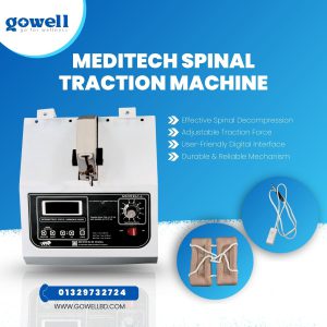
Model: Meditech Spinal Traction Machine
Modes: Intermittent, static, and harmonic modes for personalized treatment.
Interface: Digital control panel for easy adjustments.
Traction Force: Adjustable from 4 kg to 45 kg, up to 90 kg with a doubler pulley.
Timer: Adjustable treatment timer for precise sessions.
Mechanism: A Reliable and durable mechanism for consistent performance.
Treatment Areas: Spinal decompression, pain relief from herniated discs, sciatica, pinched nerves, and more.
Material: High-quality, durable construction.
Suffering from back pain, sciatica, or herniated discs? The Meditech Spinal Traction Machine provides effective, non-invasive relief by decompressing your spine and relieving pressure. With adjustable traction force, multiple modes, and a user-friendly interface, it’s perfect for pain management at home or in the clinic.


There are no reviews yet.