
Human Lumbar Spine and Pelvis Anatomical Model
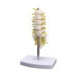
The Human Lumbar Spine and Pelvis Anatomical Model is a three-dimensional educational model designed to demonstrate the structure of the human lower spine and pelvic region.
This anatomical teaching model features representations of the five lumbar vertebrae (L1–L5), intervertebral discs, sacrum, pelvic bones, and spinal nerve roots.
The model is designed to help students and healthcare educators visualize the relationship between the lumbar spine, pelvic skeleton, and surrounding anatomical structures.
With detailed bone structures, contrasting intervertebral discs, and yellow-colored nerve representations, the model offers a practical way to study lower spinal anatomy.
A highlighted red area near the lower lumbar spine provides an additional teaching reference for discussing spinal disc abnormalities. The exact condition represented should be confirmed with the manufacturer.
The model is supported by a metal rod and a stable white display base, making it suitable for tabletop demonstrations in classrooms, medical institutions, and healthcare education environments.
Key Features and Benefits
1. Three-Dimensional Lumbar Spine Anatomy
Provides a physical representation of the lumbar spinal region, helping learners understand the structure and arrangement of the lower vertebrae.
2. Five Lumbar Vertebrae (L1–L5)
Displays the five lumbar vertebrae, which form the lower section of the vertebral column.
3. Intervertebral Disc Representation
Includes intervertebral discs positioned between the lumbar vertebrae, allowing students to visualize their anatomical arrangement.
4. Pelvic Bone Structure
Displays the pelvic bones, providing a clear visual reference for understanding the connection between the lower spine and pelvis.
5. Sacrum Representation
Features the sacrum, demonstrating its position between the lumbar spine and pelvic bones.
6. Spinal Nerve Root Details
Includes yellow-colored nerve representations extending from the vertebral region, helping learners observe the relationship between spinal bones and nerves.
7. Highlighted Spinal Disc Area
A contrasting red section near the lower lumbar region can be used as a teaching reference when discussing spinal disc conditions.
8. Educational Anatomy Demonstration
Useful for demonstrating basic lumbar spine and pelvic anatomy during medical and anatomy lessons.
9. Physiotherapy Teaching Aid
Can assist physiotherapy instructors and students in studying the structure of the lumbar spine, pelvis, and related anatomy.
10. Orthopedic and Spine Education
Provides a useful three-dimensional reference for explaining the anatomical relationship between vertebrae, discs, and pelvic structures.
11. Stable Tabletop Display Stand
Mounted on a metal support rod and white display base for convenient placement during classroom presentations.
12. Visual Learning Support
Helps students explore anatomical relationships from different viewing angles rather than relying only on printed diagrams.
Anatomical Structures Represented
- Lumbar vertebrae (L1–L5)
- Intervertebral discs
- Sacrum
- Pelvic bones
- Iliac bones
- Spinal nerve roots
- Lower spinal region
- Highlighted lumbar disc area
Applications of Human Lumbar Spine and Pelvis Anatomical Model
Medical Colleges and Universities
Suitable for anatomy lessons and medical education demonstrations.
Physiotherapy Education
Useful for explaining the relationship between lumbar vertebrae, discs, and surrounding skeletal structures.
Orthopedic Teaching
Provides a visual teaching aid for discussing basic spinal and pelvic anatomy.
Nursing and Healthcare Training
Supports introductory education about the human vertebral column and pelvic skeleton.
Anatomy Laboratories
Can be displayed as a physical reference model during practical anatomy classes.
Why Choose This Lumbar Spine and Pelvis Anatomical Model?
The Human Lumbar Spine and Pelvis Anatomical Model combines detailed lower spinal anatomy with a physical representation of the pelvic skeleton.
Its three-dimensional construction allows students to observe the arrangement of lumbar vertebrae, intervertebral discs, and nerve structures from multiple angles.
The contrasting colors of the discs and nerves make different anatomical elements easier to distinguish during teaching sessions.
Its metal support and tabletop display stand make it convenient for educational demonstrations.
Important Product Information
This anatomical model is intended for educational demonstrations and basic anatomy teaching.
It is not a diagnostic medical device and should not be used for clinical measurements, surgical planning, or patient treatment.
Exact scale, dimensions, materials, brand, detachable components, and movement capabilities should be verified with the supplier before publishing.







 No products in the cart.

Reviews
There are no reviews yet.